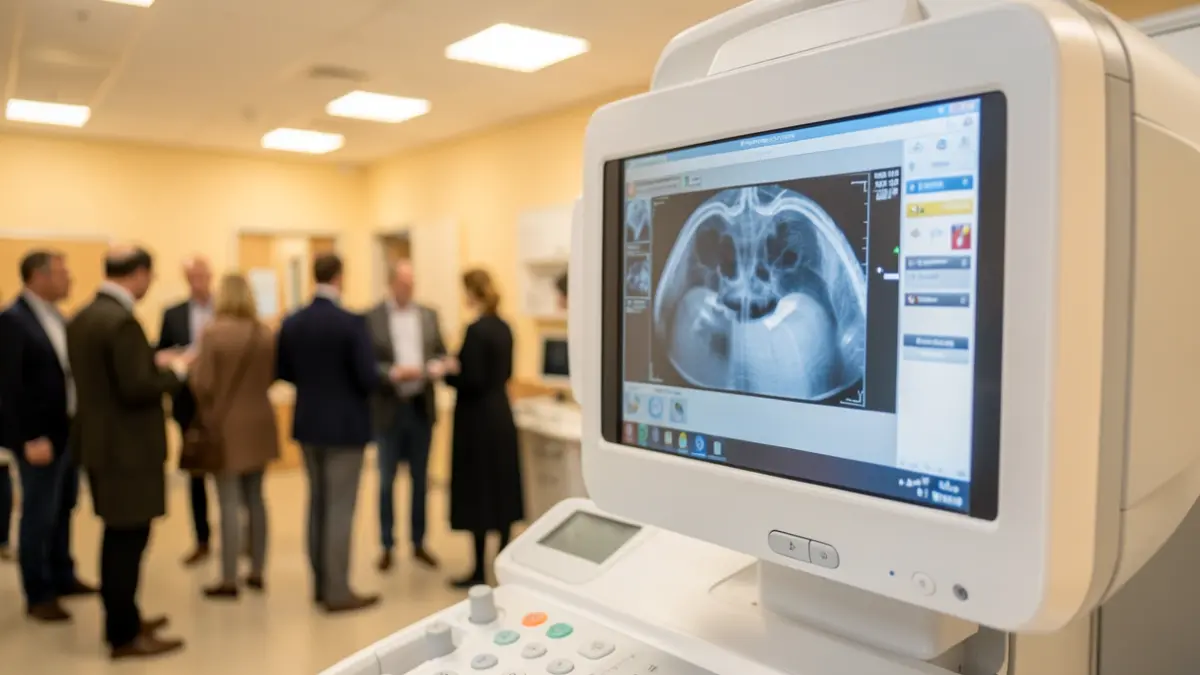
This new equipment represents a significant technological advancement, expanding the diagnostic services available for breast pathologies. It integrates various imaging techniques into a single unit, contributing to more precise, efficient, and personalized care.
The technology includes tomosynthesis (3D mammography) and artificial intelligence-based tools, which generate sharper, more detailed images. This aids in lesion detection, particularly in dense breast tissue, without increasing radiation exposure. It also facilitates image-guided biopsies and other essential procedures.
Another new feature is the capability for contrast-enhanced mammography, combining digital mammography with intravenous iodinated contrast to provide additional information on lesion vascularization.
The DUETA system allows patients to actively participate in breast compression in a controlled manner, supervised by professionals, making the examination more comfortable and reducing anxiety.
The integration of these advanced diagnostic and interventional techniques into one machine supports a more comprehensive approach to breast pathology, tailoring examinations and procedures to each patient's specific needs and reinforcing the hospital's diagnostic capacity as a leading oncological center.