
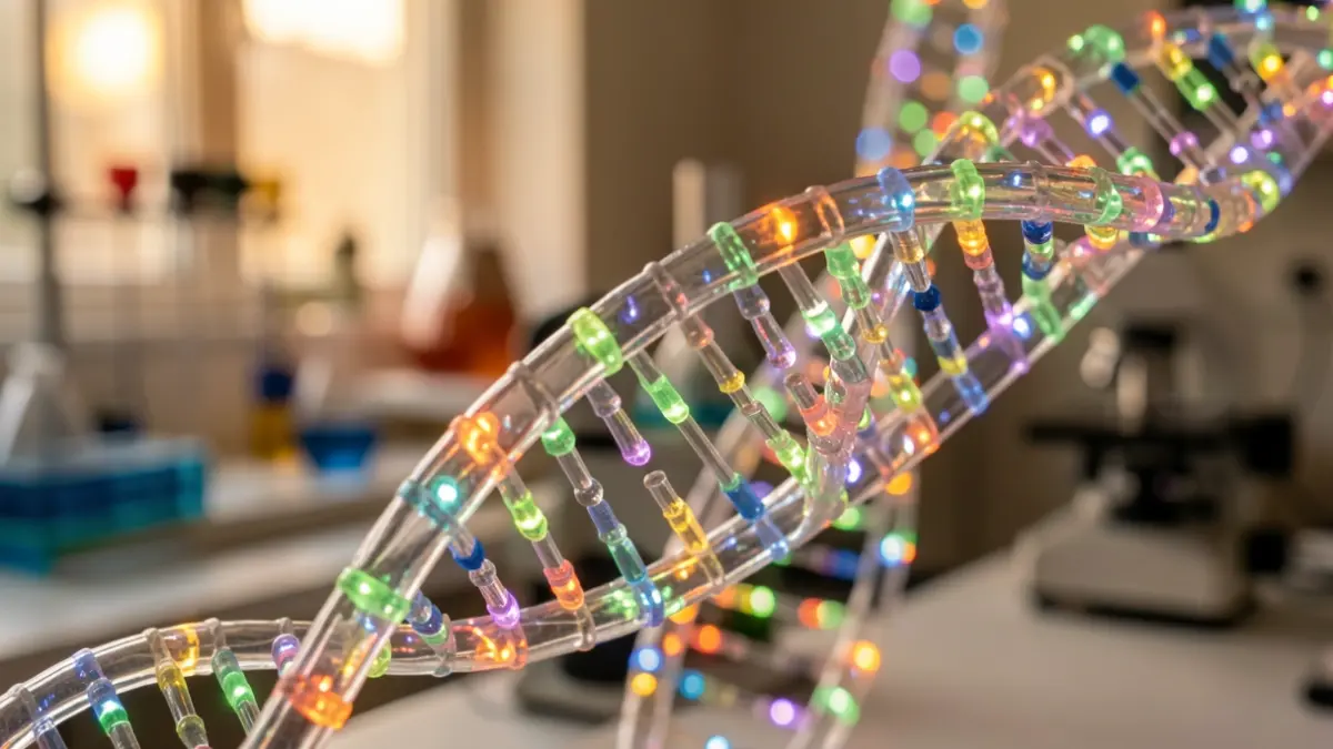
This advancement, published in the journal Molecular Cell, utilizes probes that blink intermittently, akin to 'fairy lights,' to achieve images up to ten times sharper than conventional microscopes. The technique was successfully tested on tissue samples from cancer patients, revealing that DNA in tumors is looser and more spread out than in adjacent healthy tissue.
The ability to visualize how DNA is organized in 3D within cells could become an additional tool for diagnosing and treating cancer, as DNA disorganization is linked to tumor progression. Current biopsy analysis relies on visual examination using outdated methods.
“"With the same molecule we can do two very different things. In a living cell we can watch DNA moving, which tells us how chromatin behaves. In a preserved cell we can zoom in until we are almost at the scale of the DNA molecule itself. Combining both approaches helps us see one of the main layers of control in human biology in unprecedented resolution."
The new HoT probes, designed in collaboration with researchers from City University of Hong Kong and the Guangdong Provincial People's Hospital, allow DNA visualization in living cells with 20-nanometer resolution using STORM technology. In preserved cells, precision reaches 3 nanometers with the MINFLUX technique, approaching the double helix's width (approximately 2 nanometers).
Furthermore, an artificial intelligence program named AINU has demonstrated the ability to learn from these super-resolution images. Trained on the new data, AINU correctly distinguished between skin cells and stem cells with 96-98% accuracy. This capability paves the way for future applications in cancer diagnostics and regenerative medicine.